AIMOOC
---
AIMOOC explored how machine learning could support organ-on-a-chip cultivation for personalised medicine. The wider system combined automated bright-field microscopy with TEER, oxygen and flow-rate measurements. The ML task was to turn microscope images into a useful signal about culture quality, with the longer-term goal of helping researchers monitor experiments and adjust cultivation conditions in real time.
The data problem
Biomedical imaging rarely begins with a large, balanced dataset. Our first classifier was trained on 822 microscopy images from five cell lines. Experts labelled each image as good, acceptable or bad, producing a visibly imbalanced dataset: 500 good, 212 acceptable and 110 bad samples.
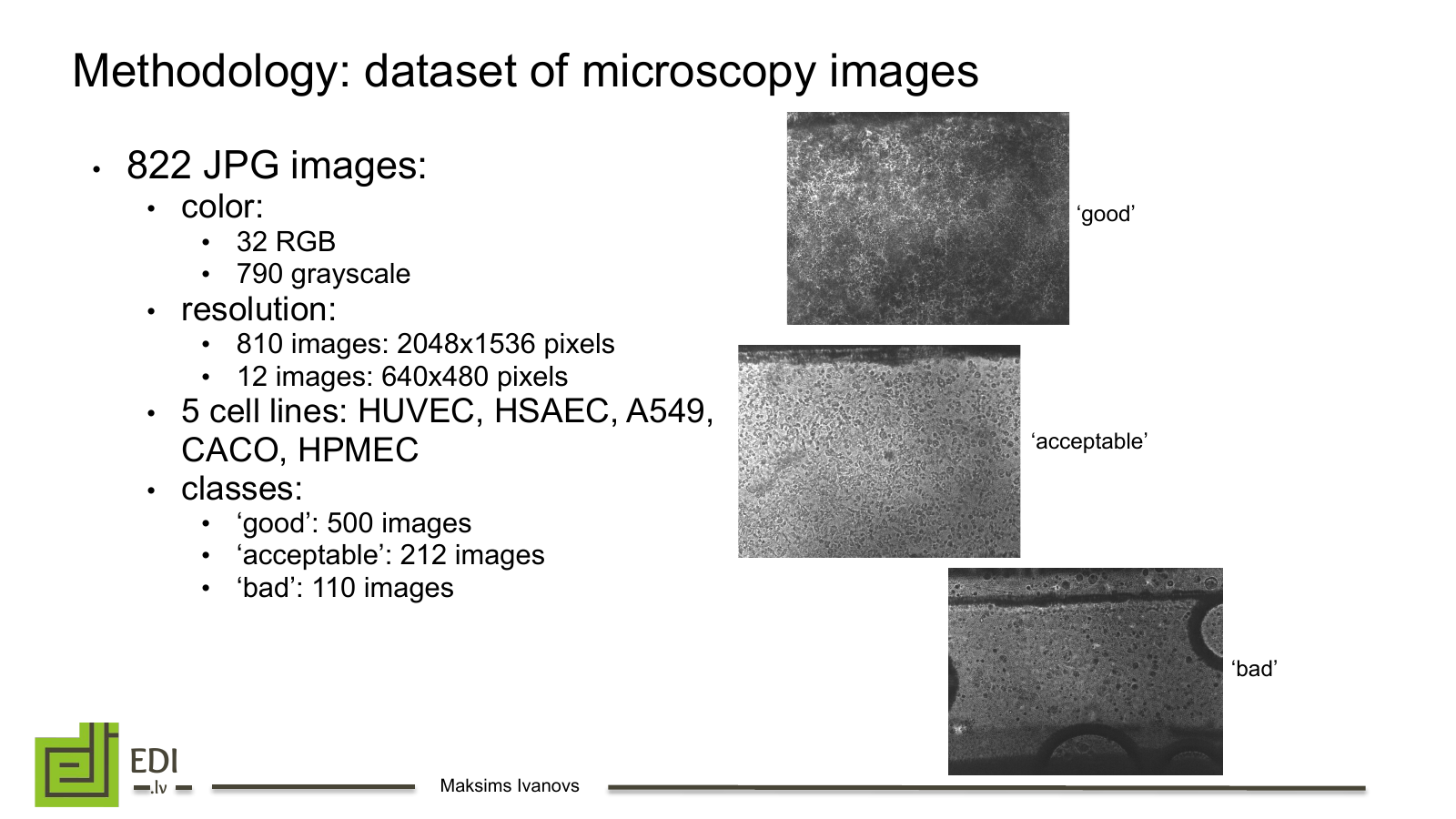
The initial microscopy dataset and its three expert-defined classes. Source: AIMOOC SPA 2023 presentation.
We divided the high-resolution images into smaller patches and used conventional augmentation to expose the model to more variation without discarding cellular detail. As data collection continued, the project produced a public dataset of 3,072 bright-field images spanning six cell lines. Its metadata includes culture quality, cell type and time after seeding, with seeding density and flow rate available for part of the collection. The dataset is split by class, cell line and time point into training, validation and test sets. Explore the OOC image dataset.
Classification
The classifier used EfficientNet-B7 pretrained on ImageNet as a frozen feature extractor. Images were resized to 600 × 600 pixels, passed through augmentation layers and the EfficientNet backbone, then through global average pooling, batch normalisation and a three-class softmax head. We trained with Adam and categorical cross-entropy, evaluating the system with five-fold cross-validation.
The best classifier reached 72.9% accuracy, compared with 60.8% for a naive majority-class baseline. This confirmed that the microscopy images contained a learnable signal, but the gap was not large enough to treat the model as a finished decision-maker—especially when moving from stable cell lines to patient-derived models.
Synthetic images
Because collecting and labelling new biological experiments is slow, we tested whether generated images could improve the classifier. We fine-tuned Stable Diffusion with LoRA on the project images, then created synthetic training sets at two LoRA weights. EfficientNet-B7 was trained on both the authentic dataset and mixtures of authentic and generated images.
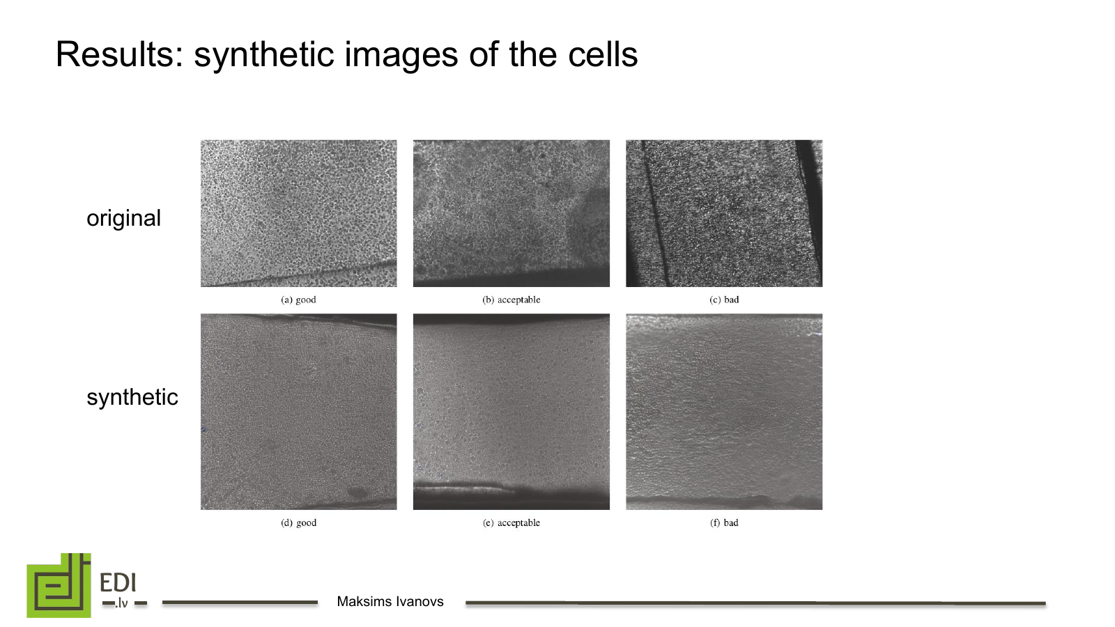
Original samples above and synthetic counterparts below. Source: AIMOOC SPA 2023 presentation.
The synthetic images resembled the source domain, but adding them made classification accuracy worse. The likely issue was not simply image quality: the generator needed to preserve class- and cell-line-specific features that mattered to the downstream task. A visually convincing cell image is not automatically a useful training example. The experiment pointed toward more controlled generation, including cell-line conditioning, better LoRA parameter selection and image-to-image translation.
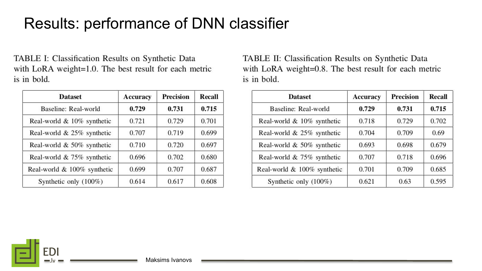
Real images produced the strongest result; increasing the synthetic share did not improve accuracy. Source: AIMOOC SPA 2023 presentation.
Outcome
The project delivered an automated bright-field imaging setup, a public OOC image dataset and an ML pipeline for assessing culture images. Tests on patient-derived iPSC lung-on-a-chip and lung-cancer-on-a-chip models also exposed the main limitation: a model trained on stable cell lines does not automatically generalise to patient material. The useful outcome was therefore both a working baseline and a clearer map of the data required for a clinically meaningful system.
AIMOOC project overview · Dataset paper · Synthetic-data paper · Conference presentation